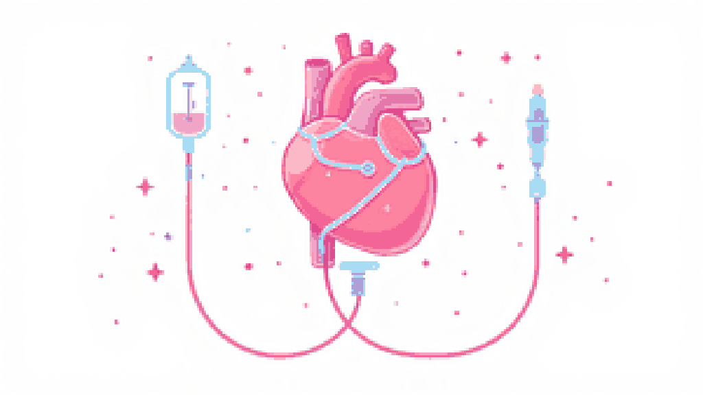
Cryoprotective perfusion is not the replacement of every water molecule in a body. It is a controlled attempt to distribute a concentrated solution through the vascular system.
The objective is to suppress ice formation during cooling while limiting osmotic stress, chemical toxicity and mechanical injury.
Why cryoprotectants are necessary
Cooling biological tissue without sufficient protection usually produces ice. Ice formation redistributes water and solutes, deforms tissue and can damage membranes and extracellular structure.
Cryoprotective agents reduce ice formation and make vitrification possible at achievable cooling rates.
Vitrification means forming an amorphous solid rather than a crystal. It is a physical state, not proof that every cell remains viable.
The modern scientific framework began with a 1984 Cryobiology paper on vitrification.
Access, washout and circulation
The surgical route depends on the protocol and the condition of the patient. Large vessels are cannulated to create an inlet and an outlet for perfusate.
A cold carrier solution is first circulated to displace blood, support pH and volume, and prepare the vasculature for cryoprotectant.
Calling this a clean canvas would be misleading. Existing clots, oedema, atherosclerosis, trauma and postmortem vascular damage remain part of the system.
Pressure, flow and temperature must be watched together. Higher pressure may improve distribution, but excessive pressure can worsen oedema or damage vessels.
Why concentration rises gradually
A sudden exposure to concentrated cryoprotectant creates large osmotic gradients. Cells can shrink rapidly, and tissue can be injured even without ice.
Protocols therefore increase concentration in stages while maintaining low temperatures. Lower temperatures generally reduce reaction rates and toxicity, but also change viscosity and flow.
Inlet and venous-effluent samples can be measured by refractometry. Convergence suggests that circulated fluid is approaching the target concentration.
It does not prove uniform concentration inside every tissue. Perfusate follows vessels, and vascular access can be uneven.
The endpoint is an estimate
Perfusion ends when protocol targets for concentration, temperature, pressure, flow and exposure time have been reached, or when further circulation is no longer useful.
The honest output is not “the patient is perfectly vitrified”. It is “the available measurements support a particular level and distribution of cryoprotection”.
Tomorrow.bio uses field cryoprotection before long-distance transport. Its public process overview says cryoprotectant is introduced gradually after stabilisation.
Its use of an optimised VM-1 formulation is also described publicly. A recent EBF research report documents continuing validation work after simulated postmortem ischaemia.
What can still go wrong
Poor vascular access can leave regions underperfused. Cryoprotectants can be toxic. Temperature and osmotic gradients can damage tissue.
These trade-offs are not evidence that perfusion is pointless. They are the reason protocol quality must be measured rather than assumed.
After perfusion, the patient proceeds to transport and a controlled cooldown through the glass transition.
Later CT imaging can test aspects of cryoprotectant distribution and ice formation, as discussed in biostasis quality checks.
TL;DR: Perfusion replaces blood and water with cryoprotective solution through the circulation. The procedure must balance ice prevention against toxicity, osmotic stress and uneven distribution.
Estimate preservation quality
Explore how timing and circumstances can change the quality of a cryopreservation procedure.
Loading the interactive tool...
Further reading